


Reviewed by
Beck Jinnette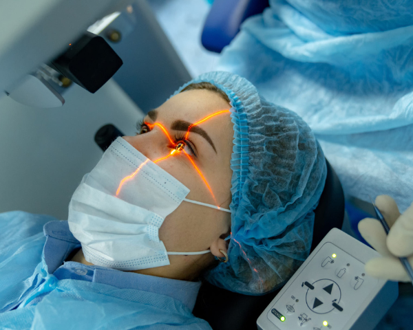
Disclaimer: We at SmartBuyGlasses are not medical doctors. This article contains general information and advice. Speak to your eye doctor to find out if you are a suitable candidate for LASIK and what kind of results you can expect.
What is LASIK eye surgery?
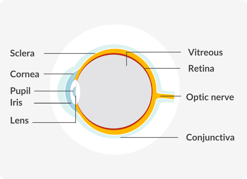
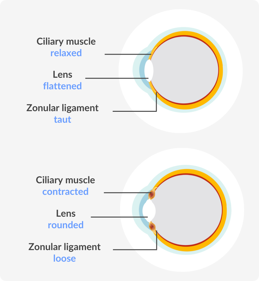
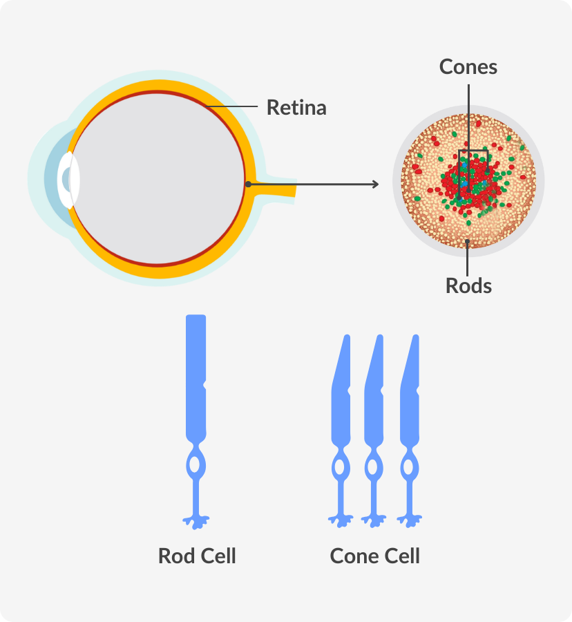
Refractive surgery is the broad term for surgical procedures that correct vision problems, and the LASIK technique is the most commonly performed one. During LASIK surgery, an excimer laser is used to reshape the transparent tissue (cornea) at the front of the eye so that light focuses directly on the retina at the back of the eye.
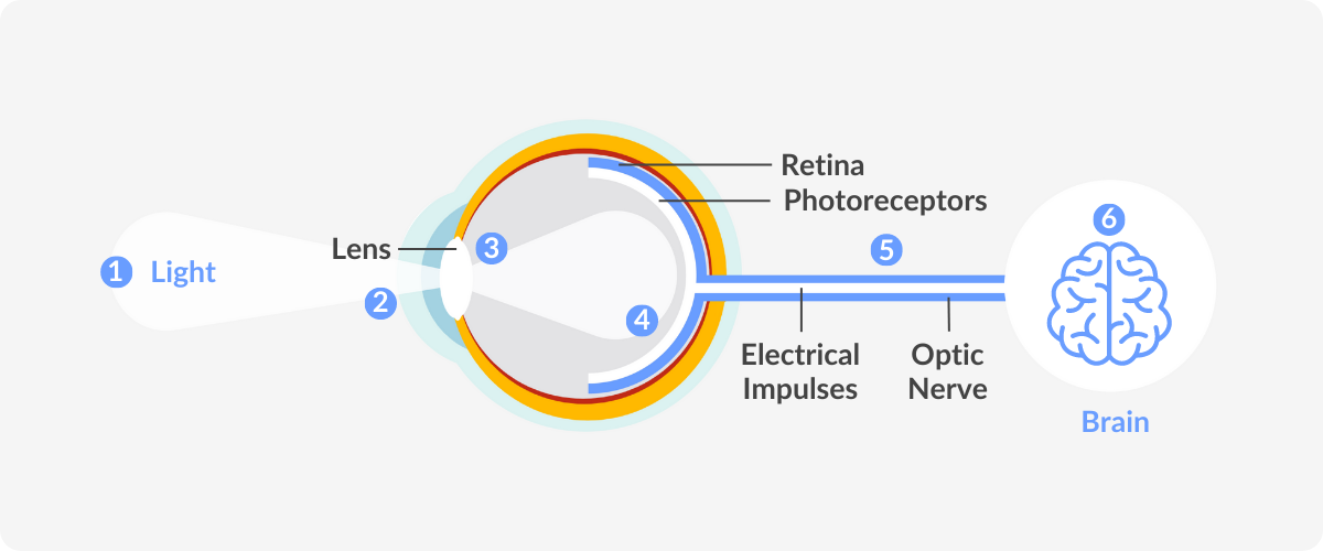
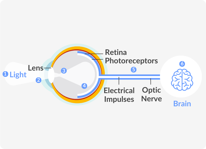
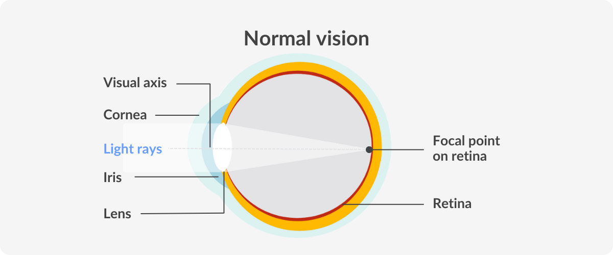
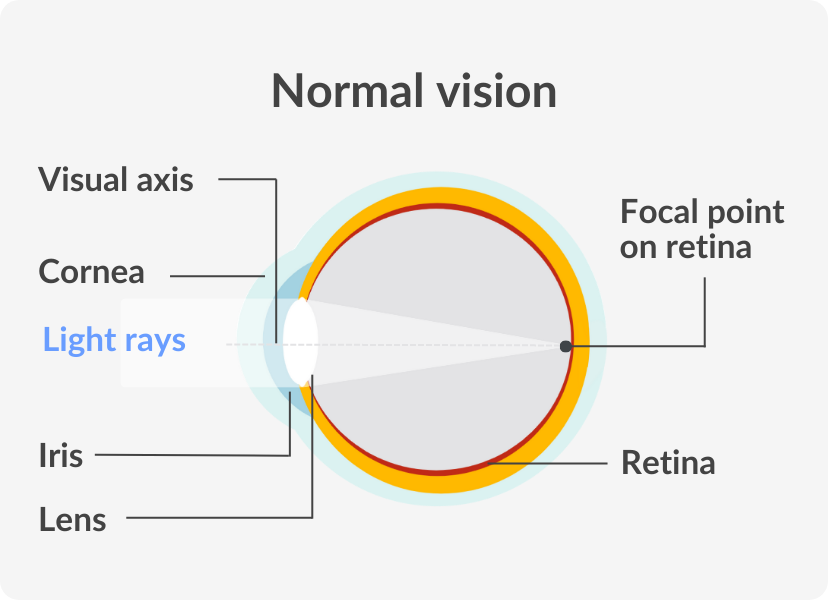
The eye refracts (bends) light as it enters the eye, and for normal vision, the focal point of the light rays must be precisely on the retina. In eyes with a refractive error such as myopia, hyperopia, or astigmatism, this is not the case, resulting in blurred vision.
LASIK eye surgery addresses this issue by modifying the shape of the cornea so that light rays are refracted correctly, thus providing clear vision. After LASIK surgery, patients can enjoy sharp visual acuity and a greater quality of life, without the need for glasses or contact lenses.
Refractive surgery is the broad term for surgical procedures that correct vision problems, and the LASIK technique is the most commonly performed one.
During LASIK surgery, an excimer laser is used to reshape the transparent tissue (cornea) at the front of the eye so that light focuses directly on the retina at the back of the eye.
The eye refracts (bends) light as it enters the eye, and for normal vision, the focal point of the light rays must be precisely on the retina.
In eyes with a refractive error such as myopia, hyperopia, or astigmatism, this is not the case, resulting in blurred vision.
LASIK eye surgery addresses this issue by modifying the shape of the cornea so that light rays are refracted correctly, thus providing clear vision.
After LASIK surgery, patients can enjoy sharp visual acuity and a greater quality of life, without the need for glasses or contact lenses.
What conditions can LASIK eye surgery treat?
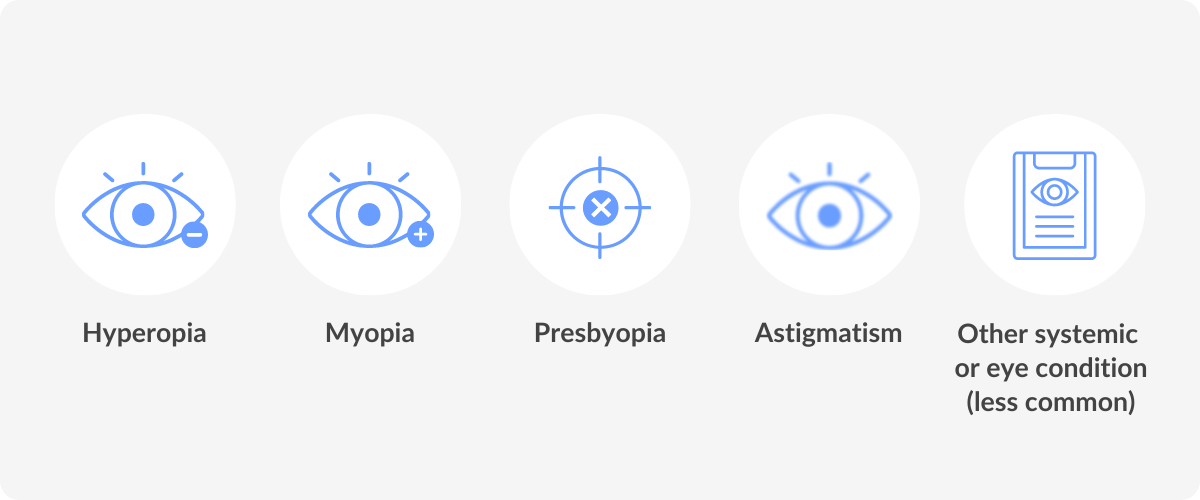
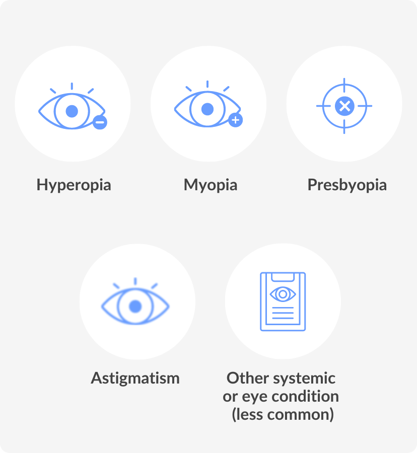
LASIK surgery is used to treat refractive errors, which are the most common causes of vision problems. The three most common refractive errors are myopia, hyperopia and astigmatism.
In each case, light is refracted incorrectly but in different ways. If you have one of these conditions, refractive eye surgery may be effective in improving vision, as long as it falls within the treatment limits.
Myopia
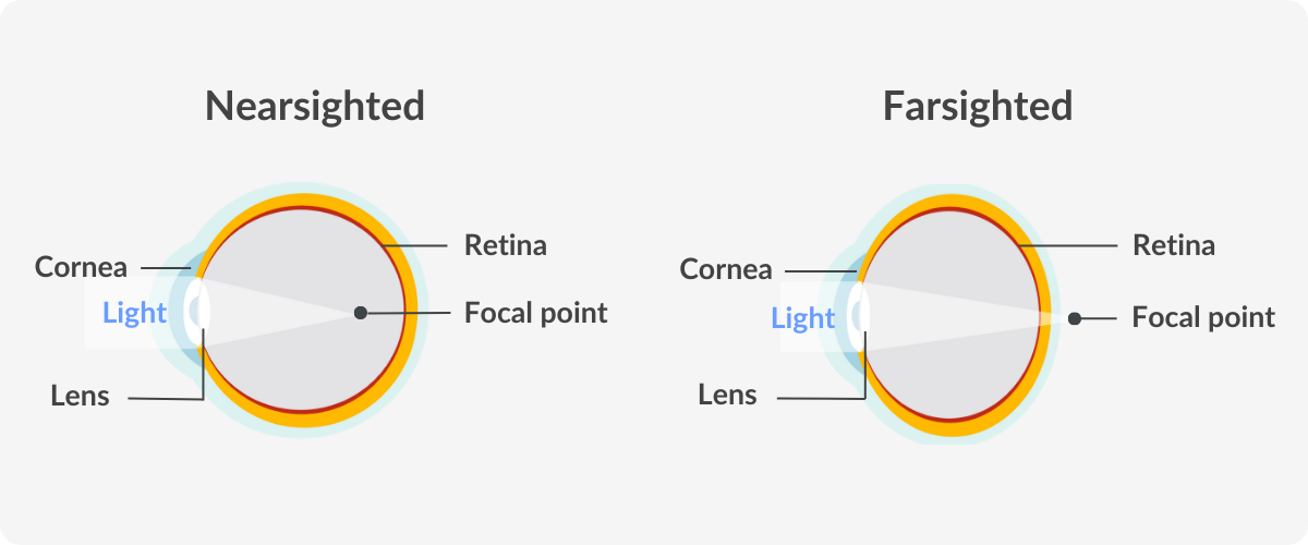
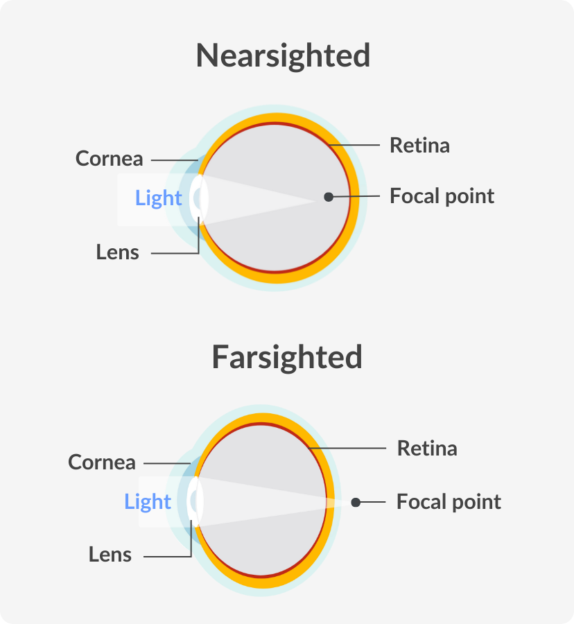
Myopia, also known as shortsightedness and nearsightedness, is the most common vision problem in the world, affecting about 33% of the global population. People with shortsightedness have no problem with their close-up vision, but experience blurred vision when looking at distant objects.
With a nearsighted eye, the eyeball itself is elongated, causing the focal point of the light rays entering the eye to fall short of the retina. An overly-curved cornea can also have this effect, with the same vision results.
Myopia, also known as shortsightedness and nearsightedness, is the most common vision problem in the world, affecting about 33% of the global population.
People with shortsightedness have no problem with their close-up vision, but experience blurred vision when looking at distant objects.
With a nearsighted eye, the eyeball itself is elongated, causing the focal point of the light rays entering the eye to fall short of the retina.
An overly-curved cornea can also have this effect, with the same vision results.
Hyperopia
Hyperopia is commonly referred to as farsightedness, and, as the name suggests, is the inverse of nearsightedness. People with hyperopia have clear distance vision, but nearby objects appear fuzzy or blurry. An eyeball that is short in length is the cause of hyperopia, focusing light rays beyond the retina.
Hyperopia is commonly referred to as farsightedness, and, as the name suggests, is the inverse of nearsightedness. People with hyperopia have clear distance vision, but nearby objects appear fuzzy or blurry.
An eyeball that is short in length is the cause of hyperopia, focusing light rays beyond the retina.
Astigmatism
People affected by this refractive error have blurry vision both up close and farther away. An irregularly shaped cornea or lens is responsible for astigmatism.
Does LASIK eye surgery permanently correct your vision?
LASIK surgery alters the shape of your cornea, based on your vision requirements at the time of your surgery. This is a permanent change, and the corneal tissue will never revert to its previous shape. However, due to the natural aging process of the eyes, your vision will likely begin to deteriorate at some point, possibly necessitating glasses again.
Presbyopia is a condition affecting close-distance vision, and it tends to develop in most adults from the age of 40 onwards, again, as a natural part of aging. Even after a LASIK procedure, you’ll probably need to use reading glasses at some stage to treat presbyopia, but this will likely be years later.
Vision changes that occur after having refractive surgery are not an indication that the procedure has not worked, but a consequence of unrelated conditions that may develop afterwards. Many patients enjoy consistent vision for ten years or more following surgery (Ide et al., 2014).
LASIK surgery alters the shape of your cornea, based on your vision requirements at the time of your surgery.
This is a permanent change, and the corneal tissue will never revert to its previous shape. However, due to the natural aging process of the eyes, your vision will likely begin to deteriorate at some point, possibly necessitating glasses again.
Presbyopia is a condition affecting close-distance vision, and it tends to develop in most adults from the age of 40 onwards, again, as a natural part of aging.
Even after a LASIK procedure, you’ll probably need to use reading glasses at some stage to treat presbyopia, but this will likely be years later.
Vision changes that occur after having refractive surgery are not an indication that the procedure has not worked, but a consequence of unrelated conditions that may develop afterwards.
Many patients enjoy consistent vision for ten years or more following surgery (Ide et al., 2014).
Who is a good candidate for LASIK eye surgery?
Laser vision correction is not a suitable option for everyone. Even if you have a refractive error, there is no guarantee that the specifics of your condition will make it a viable option for you. You must also meet the following criteria in order to have the procedure:
- You must be at least 18 years old, although it is better to wait until you’re in your 20s when your vision is more likely to have stabilised
- Your vision prescription should have remained constant for at least the last year
- Your corneas must be thick and healthy, and you must have healthy eyes in general
- You need to have realistic expectations about what LASIK can do for your vision
Laser vision correction is not a suitable option for everyone. Even if you have a refractive error, there is no guarantee that the specifics of your condition will make it a viable option for you.
You must also meet the following criteria in order to have the procedure:
- You must be at least 18 years old, although it is better to wait until you’re in your 20s when your vision is more likely to have stabilised
- Your vision prescription should have remained constant for at least the last year
- Your corneas must be thick and healthy, and you must have healthy eyes in general
- You need to have realistic expectations about what LASIK can do for your vision
Unsuitable candidates
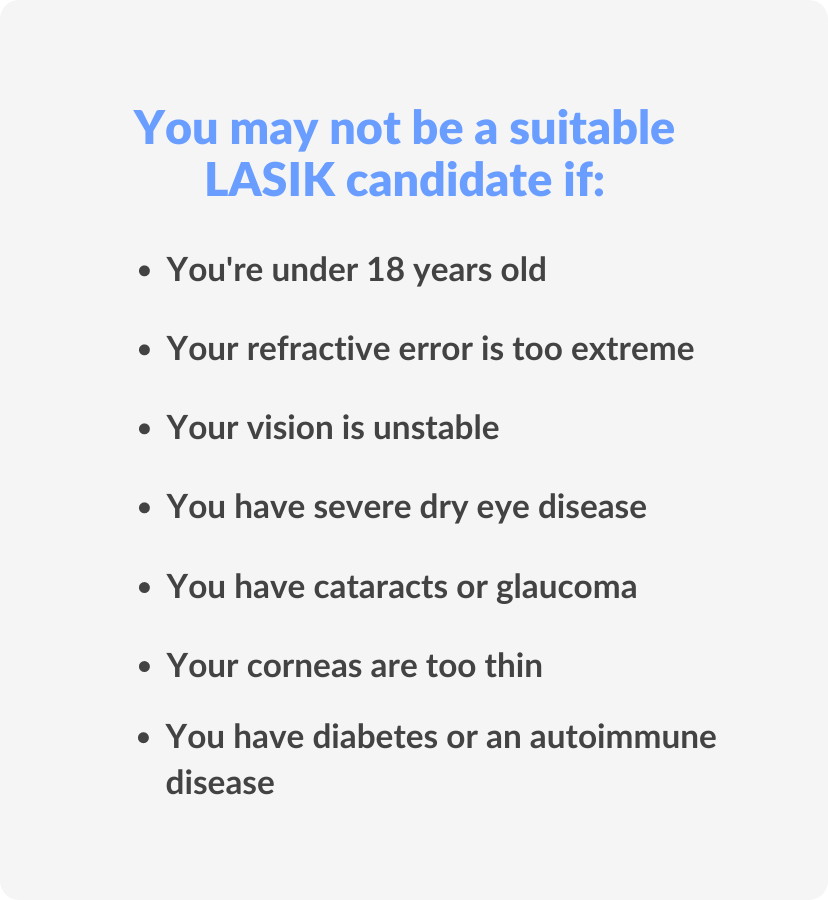
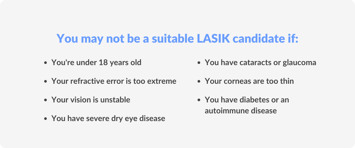
Even if you satisfy the requirements mentioned above, there are, unfortunately, some factors that can directly rule out the possibility of undergoing LASIK surgery. If any of the following apply to you, LASIK may not be a feasible option:
- Your refractive error is unstable (still worsening)
- You have extreme myopia, hyperopia, or astigmatism
- You suffer from severe dry eye syndrome
- You have overly thin corneas
- You have scars on your corneal tissue
- You have keratoconus (cone-shaped corneas)
- You have advanced glaucoma
- You have cataracts
- You have a history of certain eye conditions
- You have diabetes that is not well controlled
Even if you satisfy the requirements mentioned above, there are, unfortunately, some factors that can directly rule out the possibility of undergoing LASIK surgery.
If any of the following apply to you, LASIK may not be a feasible option:
- Your refractive error is unstable (still worsening)
- You have extreme myopia, hyperopia, or astigmatism
- You suffer from severe dry eye syndrome
- You have overly thin corneas
- You have scars on your corneal tissue
- You have keratoconus (cone-shaped corneas)
- You have advanced glaucoma
- You have cataracts
- You have a history of certain eye conditions
- You have diabetes that is not well controlled
Preparation for LASIK eye surgery
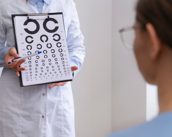
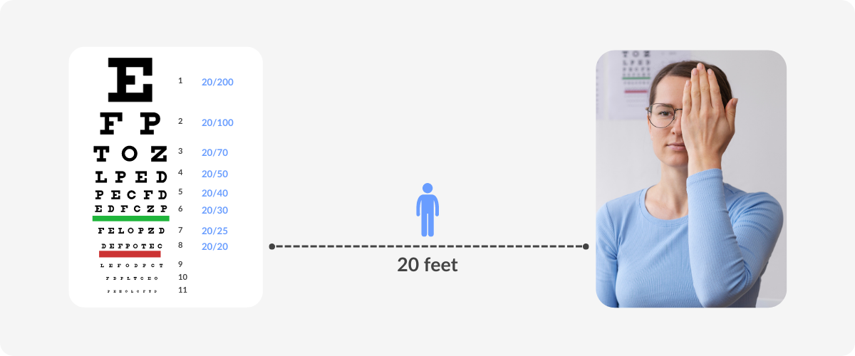
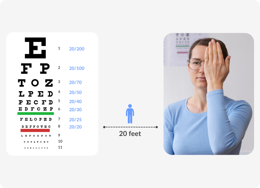
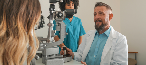
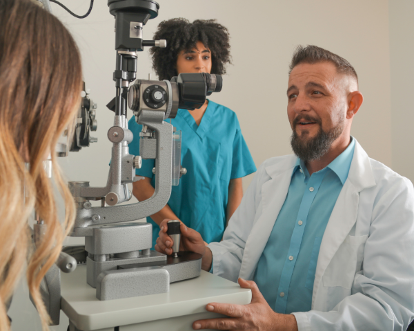
The first thing you need to do when considering LASIK surgery is to speak with an opthalmologist. They will be able to tell you to what extent the surgical procedure could be effective in your case, and give you realistic expectations for the outcome. They will then test your vision thoroughly to ensure you are a good candidate.
For those who wear contact lenses, you’ll have to take a break from them and wear glasses only for a few weeks before the eye doctor evaluates your potential as a candidate. The exact duration will depend on the type of contact lenses you wear and how long you’ve been using them. Your eye doctor will give you precise guidelines for this.
The opthalmologist will then check for the presence of other eye problems by means of a comprehensive eye exam. If there are no complications, they will then map the surface of your cornea (corneal topography) and check its thickness, as well as measure your pupils. These details are important for programming the computer used for laser surgery.
The first thing you need to do when considering LASIK surgery is to speak with an opthalmologist.
They will be able to tell you to what extent the surgical procedure could be effective in your case, and give you realistic expectations for the outcome.
They will then test your vision thoroughly to ensure you are a good candidate.
For those who wear contact lenses, you’ll have to take a break from them and wear glasses only for a few weeks before the eye doctor evaluates your potential as a candidate.
The exact duration will depend on the type of contact lenses you wear and how long you’ve been using them. Your eye doctor will give you precise guidelines for this.
The opthalmologist will then check for the presence of other eye problems by means of a comprehensive eye exam.
If there are no complications, they will then map the surface of your cornea (corneal topography) and check its thickness, as well as measure your pupils.
These details are important for programming the computer used for laser surgery.
The procedure
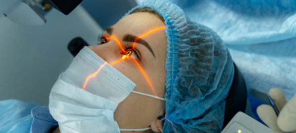
During laser eye surgery, the ophthalmologist or refractive surgeon will use eye drops to numb your eyes. They will then place a suction ring and eyelid speculum in your eye. These devices do not cause any pain, but you may feel some pressure from them. They hold your eyes in the correct position while also preventing you from blinking.
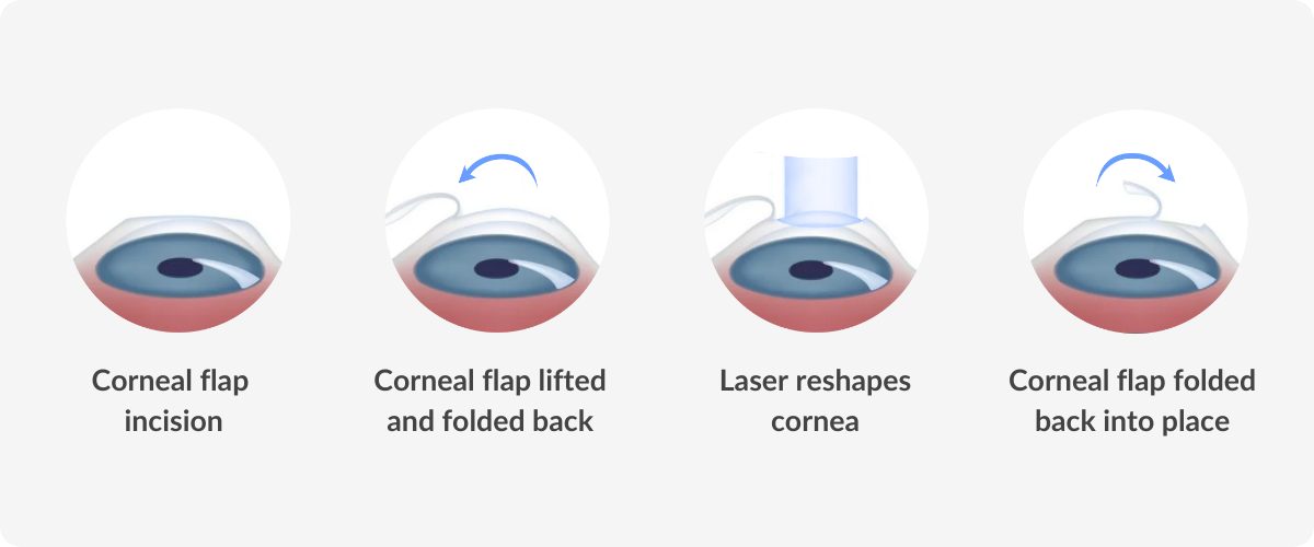
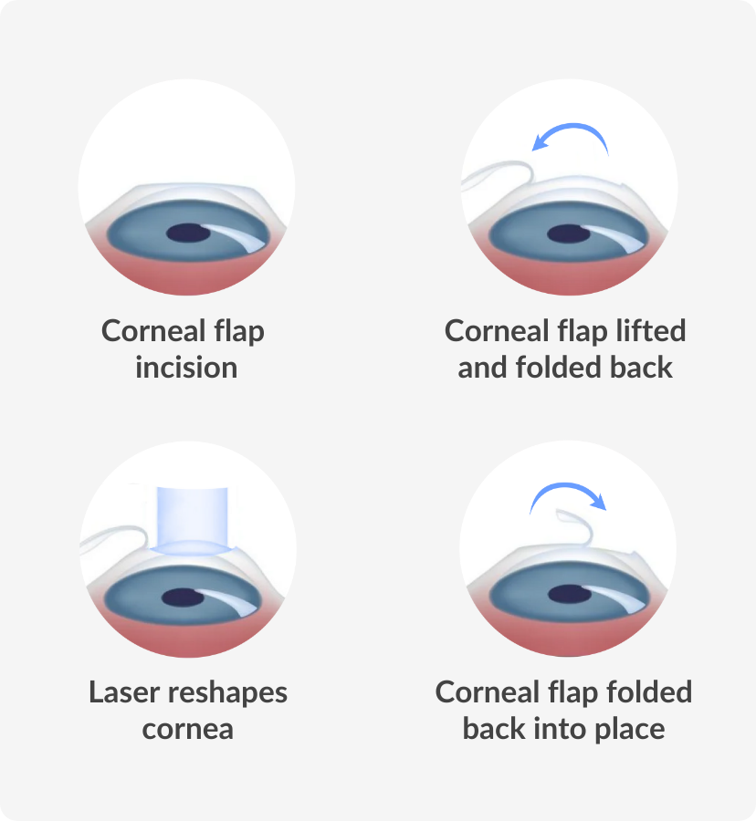
Using a laser or a device called a microkeratome, the eye surgeon will make an incision in your corneal tissue to create a thin flap, which is then folded back. You will be asked to stare at a fixed point ahead of you while the eye surgeon uses the laser to reshape your cornea according to the measurements taken by your ophthalmologist.
During this part of the procedure, you will hear a clicking sound and notice a strange odour. This is no cause for alarm, just a normal part of the laser treatment. The smell is created by the chemical reaction from the laser beam shaping the cornea.
Once the reshaping is complete, the eye surgeon will fold the corneal flap back over to its original position, where it will reattach by itself within two to three minutes and begin to heal.
During laser eye surgery, the ophthalmologist or refractive surgeon will use eye drops to numb your eyes. They will then place a suction ring and eyelid speculum in your eye.
These devices do not cause any pain, but you may feel some pressure from them. They hold your eyes in the correct position while also preventing you from blinking.
Using a laser or a device called a microkeratome, the eye surgeon will make an incision in your corneal tissue to create a thin flap, which is then folded back.
You will be asked to stare at a fixed point ahead of you while the eye surgeon uses the laser to reshape your cornea according to the measurements taken by your ophthalmologist.
During this part of the procedure, you will hear a clicking sound and notice a strange odour. This is no cause for alarm, just a normal part of the laser treatment.
The smell is created by the chemical reaction from the laser beam shaping the cornea.
Once the reshaping is complete, the eye surgeon will fold the corneal flap back over to its original position, where it will reattach by itself within two to three minutes and begin to heal.
Aftercare
You’ll be able to see after surgery, and your old corrective lenses won’t be effective anymore, but it will take two to three months for your vision to fully sharpen and stabilise. Patients usually experience a gritty, itchy, or burning sensation in their eyes immediately after laser surgery. You may be given eye drops or pain medication to use in the hours and days that follow.
You’ll have a follow-up appointment with your eye surgeon within a day or two of your surgery. They will check your condition and give you precise recommendations which must be closely followed throughout the healing process.
You’ll receive a protective shield to wear on your eyes while sleeping to avoid unwanted contact and prevent infection. This can be used for several weeks until the eyes heal. It’s essential to refrain from touching your eyes even in the daytime to allow the corneal flap to heal correctly.
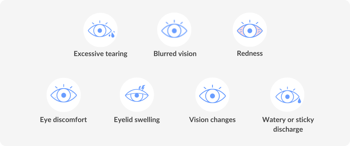
You may have some pain, dry or watery eyes, puffy eyelids, or increased light sensitivity in the first week or so after laser eye surgery. You may also see a halo around bright lights for a while. This is normal and will subside with time. Eye drops can be effective in relieving dry eyes, but if you feel severe pain or that these symptoms are not improving as they should, consult your eye doctor.
Some activities pose risks to the healing process, so you may have to modify your regular routine for a little while after your procedure. It’s generally advised to avoid exercise for three days and refrain from playing contact sports for the first month post-op. Don’t use eye makeup or cosmetic products on or near the eyes for two weeks, and stay away from hot tubs for two months.
If you have doubts about any other activities that are a regular part of your lifestyle, ask your eye doctor’s advice and follow the time guidelines they give you.
You’ll be able to see after surgery, and your old corrective lenses won’t be effective anymore, but it will take two to three months for your vision to fully sharpen and stabilise.
Patients usually experience a gritty, itchy, or burning sensation in their eyes immediately after laser surgery. You may be given eye drops or pain medication to use in the hours and days that follow.
You’ll have a follow-up appointment with your eye surgeon within a day or two of your surgery.
They will check your condition and give you precise recommendations which must be closely followed throughout the healing process.
You’ll receive a protective shield to wear on your eyes while sleeping to avoid unwanted contact and prevent infection. This can be used for several weeks until the eyes heal.
It’s essential to refrain from touching your eyes even in the daytime to allow the corneal flap to heal correctly.
You may have some pain, dry or watery eyes, puffy eyelids, or increased light sensitivity in the first week or so after laser eye surgery.
You may also see a halo around bright lights for a while. This is normal and will subside with time.
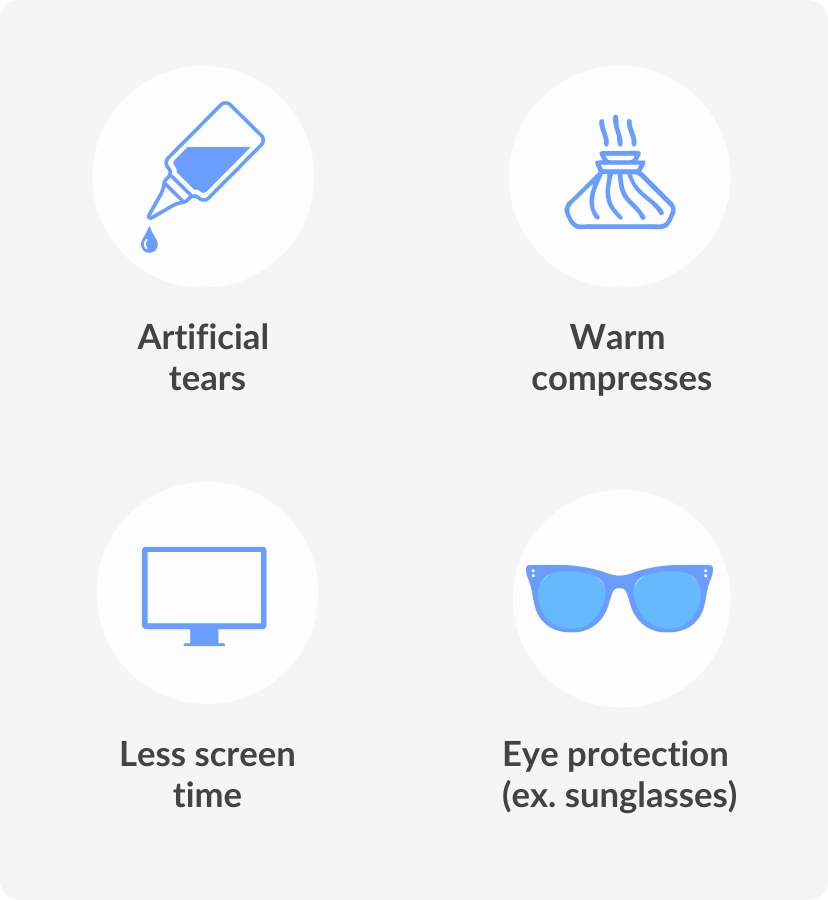
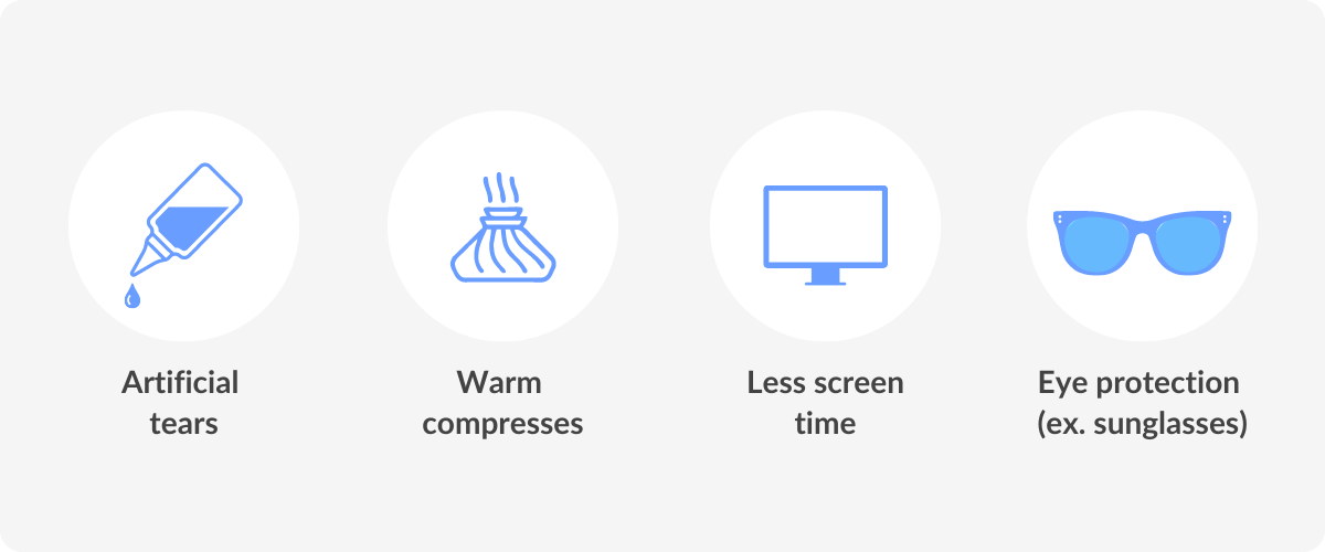
Eye drops can be effective in relieving dry eyes, but if you feel severe pain or that these symptoms are not improving as they should, consult your eye doctor.
Some activities pose risks to the healing process, so you may have to modify your regular routine for a little while after your procedure.
It’s generally advised to avoid exercise for three days and refrain from playing contact sports for the first month post-op.
Don’t use eye makeup or cosmetic products on or near the eyes for two weeks, and stay away from hot tubs for two months.
If you have doubts about any other activities that are a regular part of your lifestyle, ask your eye doctor’s advice and follow the time guidelines they give you.
DID YOU KNOW?
The procedure usually only takes about 5 minutes per eye, with the actual laser treatment generally taking 5-15 seconds per eye.
What are the risks and side effects associated with LASIK?
There are risks associated with every kind of surgery, and refractive surgery is no different. The initial side effects mentioned in the section above generally go away after a week or two, but in some cases, they may continue for longer. Changing vision throughout the day is another initial side effect that can persist for some people.
Apart from standard side effects, there is the risk of complications stemming from laser eye surgery. Over 99% of patients do not experience any, according to the Cleveland Clinic, but it is useful to be aware of them.
- Issues could arise with the cornea, potentially requiring treatment or surgery
- Inflammation or infection of the eye could occur, which can be treated with medication
- Eyesight that is worse than your pre-LASIK vision, usually untreatable
It is very rare that LASIK patients experience vision loss as a result of refractive surgery, but it is still possible. The risk of any surgical complications occurring with LASIK is less than 1%. There is always the risk that you may be disappointed with the results, especially if your expectations are too high, or if you’re hoping to never again have to wear contact lenses or glasses.
There are risks associated with every kind of surgery, and refractive surgery is no different.
The initial side effects mentioned in the section above generally go away after a week or two, but in some cases, they may continue for longer.
Changing vision throughout the day is another initial side effect that can persist for some people.
Apart from standard side effects, there is the risk of complications stemming from laser eye surgery.
Over 99% of patients do not experience any, according to the Cleveland Clinic, but it is useful to be aware of them.
- Issues could arise with the cornea, potentially requiring treatment or surgery
- Inflammation or infection of the eye could occur, which can be treated with medication
- Eyesight that is worse than your pre-LASIK vision, usually untreatable
It is very rare that LASIK patients experience vision loss as a result of refractive surgery, but it is still possible. The risk of any surgical complications occurring with LASIK is less than 1%.
There is always the risk that you may be disappointed with the results, especially if your expectations are too high, or if you’re hoping to never again have to wear contact lenses or glasses.
Is LASIK eye surgery right for me?
Refractive surgery can be a life-changing procedure, allowing people to live more freely without having to worry about glasses or contact lenses. Many activities become much easier and more comfortable, such as water and contact sports, as well as the simple fact of waking up in the morning and being able to see immediately, without having to reach for your glasses or contact lenses.
As outlined in this article, having laser eye surgery is not simply a matter of deciding whether you want it or not. There are many criteria that must be met in terms of your eye and personal health, as well as the changes it will have to your personal life during the recovery period, and of course, the financial aspect.
Speak to your eye doctor about laser refractive surgery; they will be able to talk to you in detail about your concerns, and have the relevant knowledge of your existing medical conditions, your medical history and how it could be beneficial for you personally. In the meantime, feel free to ask our Opticians if you have any other enquiries about LASIK surgery or general eye health.
Refractive surgery can be a life-changing procedure, allowing people to live more freely without having to worry about glasses or contact lenses.
Many activities become much easier and more comfortable, such as water and contact sports, as well as the simple fact of waking up in the morning and being able to see immediately, without having to reach for your glasses or contact lenses.
As outlined in this article, having laser eye surgery is not simply a matter of deciding whether you want it or not.
There are many criteria that must be met in terms of your eye and personal health, as well as the changes it will have to your personal life during the recovery period, and of course, the financial aspect.
Speak to your eye doctor about laser refractive surgery; they will be able to talk to you in detail about your concerns, and have the relevant knowledge of your existing medical conditions, your medical history, and how it could be beneficial for you personally.
In the meantime, feel free to ask our Opticians if you have any other inquiries about LASIK surgery or general eye health.
References
Schallhorn et al. (2016), Patient-reported outcomes 5 years after laser in situ keratomileusis, Journal of Cataract & Refractive Surgery https://journals.lww.com/jcrs/Abstract/2016/06000
Ide et al. (2014), Outcome of a 10-year follow-up of laser in situ laser keratomileusis for myopia and myopic astigmatism, Taiwan Journal of Ophthalmology https://www.sciencedirect.com/science/article/pii/via%3Dihub
Cleveland Clinic, LASIK Eye Surgery. https://my.clevelandclinic.org/health/treatments/21805-lasik-eye-surgery
Mayo Clinic, LASIK Eye Surgery.Cleveland Clinic, LASIK Eye Surgery. https://www.mayoclinic.org/tests-procedures/lasik-eye-surgery/about/pac-20384774